Multiple Sclerosis Lesion Segmentation on 7T MRI: A U-Net Tool and Evaluation
The recent introduction of 7T MRI to multiple sclerosis has improved characterization of lesional pathology, particularly through increased resolution.
However, due to unique signal characteristics and scanner artifacts, existing methods for automatic lesion detection do not translate well to 7T MRI. For this reason, no standardized, universal, and well validated tool for automated 7T MRI MS lesion segmentation exists.
Existing work on MS lesion detection in 7T scans includes work with MP2RAGE acquisitions, [1, 2] despite the more typical use of FLAIR for lesion quantification in MS research.
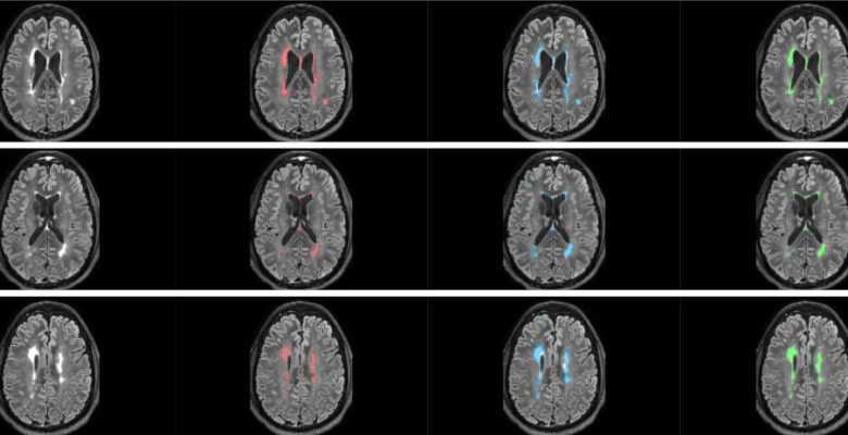
Here we explore state-of-the-art methods in Deep Learning applied to the task of 7T white matter lesion (WML) segmentation on FLAIR, with plans to provide a tool for 7T WML to the MS research community.
